
محققان فقط با تحریک بخشیهایی از مغز یک مرد، موفق به انتقال مجازی وی به زمان گذشته شدند. یک مرد 22 ساله در این تجربه، تماشای صحنههایی از خانوادهاش و همچنین ایستگاه قطار محلی را حین نشستن در اتاق پزشکی گزارش کرد. محققان امیدوارند این دستاورد بتواند به آنها در درک بهتر نواحی مغزی که برای بازیابی اطلاعات درباره مکانها به کار میرود، کمک کند.
این توهمات، زمانی که «پیر مگهوند» از «موسسه فاینستاین» در نیویورک الکترودهایی را در درون مغز یک مرد جوان با هدف جستجوی منشا صرع وی کاشت، ظاهر شدند. این امر شامل حفاری روزنههای ریز در جمجمه وی بود که از طریق آن، پزشکان دو الکترود پنج سانتیمتری را وارد کرده و آنها را به نقاط خاصی از بافت مغز هدایت کردند.
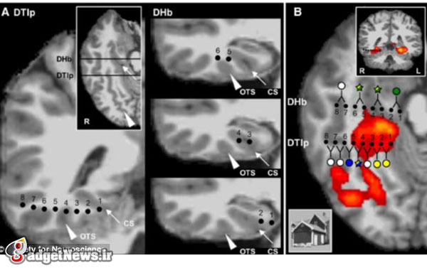
دکتر مگهوند نخست با استفاده از امآرآی کاربردی و زمانی که بیمار به تصاویری از اشیا و صحنههای مختلف نگاه میکرد، از مغز وی اسکن گرفت. سپس فعالیت الکترودهای کاشتشده زمانی که وی به مجموعهای مشابه از تصاویر نگاه میکرد، ضبط شد.
تیم علمی دریافت ناحیهای از قشر مغزی در اطراف هیپوکامپ زمانی که سوژه به تصاویری از مکانها نگاه میکرد، فعال میشد. این نقاط ریز بافتهایی بودند که به نظر میرسید به خانهها و مکانها بیش از دیگر اشیا اهمیت میدهند. الکترودهای کاشتهشده برای تحریک مغز در این ناحیه به کار رفتند و مجموعهای از توهمات بصری تولید شدند.
مرد جوان در ابتدا تماشای یک ایستگاه راهآهن آشنا و سپس بخشی از منزلش را گزارش کرد، اما هیچ بو یا صدایی با این صحنهها مرتبط نبود. زمانی که تحریک این نواحی مغزی تکرار شد، همان توهمات اتفاق افتاد. هنگامی که محققان الکترودها را در بخشی اندکی متفاوت و در شکنج گیجگاهی تحتانی تحریک کردند، داوطلب عنوان کرد که چهرهها به ناگاه از ریخت افتادند.
آشش مهتا، یکی از اعضای تیم علمی، در این باره گفت: شاید گروه خاصی از عصبها، خاطره فردی که پیشبند به تن دارد را کدبندی کرده و گروه دیگر اجاق یا یک خیابان را رمزگذاری میکنند و زمانی که آنها به همراه یکدیگر تحریک میشوند، خاطره آشنایی از آن مکان را به همراه تمامی اشیا در آنجا تداعی میکنند.
مهتا معتقد است این آزمایشها شاید در نهایت به افرادی کمک کند که از شرایطی مانند اوتیسم یا بیماری آلزایمر رنج میبرند.
منبع : easybranches
Forget virtual reality headsets: Doctors transport a patient to his past by zapping the brain to make him HALLUCINATE
Patient said he saw scenes from his family's pizzeria while in the hospital
The hallucinations appeared after electrodes activated regions in his brain
Area around hippocampus was found to be involved in the hallucination
The findings may eventually help develop treatments for people who suffer from conditions such as autism or Alzheimer's disease
Scientists have been able to virtually transport a man back in time – simply by stimulating parts of his brain.
A 22-year-old man reported seeing scenes from his family’s pizzeria as well as his local train station, all while sitting in a medical room.
Researchers hope the discovery could help them better understand areas of the brain used to retrieve information on locations.
The black dots represent the probes researchers used to cause a hallucination in their patient. Dr Mégevand at the Feinstein Institute in New York first scanned the volunteer's brain using functional MRI while he looked at images of different objects and scenes. Activity from the implanted electrodes was then recorded as the patient looked at a similar set of pictures
The black dots represent the probes researchers used to cause a hallucination in their patient. Dr Mégevand at the Feinstein Institute in New York first scanned the volunteer's brain using functional MRI while he looked at images of different objects and scenes. Activity from the implanted electrodes was then recorded as the patient looked at a similar set of pictures
The halluciations appeared after Pierre Mégevand at the Feinstein Institute in New York implanted electrodes into the young man’s brain in search of the origin of his epilepsy.
This involved drilling tiny holes in the skull through which doctors inserted 2 inch-long (5cm) electrodes and guided them to specific points in the brain tissue.
Dr Mégevand first scanned the volunteer's brain using functional MRI while he looked at images of different objects and scenes, according to a report by Helen Thomson in New Scientist.
The team found that an area of the cortex around the hippocampus was activated when the subject looked at images of places.
‘There are these little spots of tissues that seem to care about houses and places more than any other class of object,’ team member Ashesh Mehta at the Feinstein Institute told the New Scientist.
The implanted electrodes were used to stimulate the brain in this area which triggered a series of visual hallucinations.
The young man initially described seeing a familiar railway station and later a part of his home, but no smells or sounds were associated with the scenes.
When stimulation of these brain areas was repeated, the same hallucinations occurred.
And when the researchers stimulated electrodes in a slightly different area, in the inferior temporal gyrus (ITG), the volunteer said that faces suddenly became distorted.
‘Maybe a specific group of neurons encodes a memory of a person wearing an apron, another group encodes an oven or a street, and when you stimulate them altogether it evokes a familiar memory of that place where all those things were,’ said Dr Mehta.
Dr Mehta said the experiments may eventually help people who who suffer from conditions such as autism or Alzheimer's disease.
The research follows a study at Stanford University, which two years ago revealed that electrodes placed in another area of the brain could change a person’s ability to process faces.
When the researchers stimulated electrodes in a slightly different area, in the inferior temporal gyrus (ITG) shown here in yellow, the volunteer said that the doctor's faces suddenly became distorted